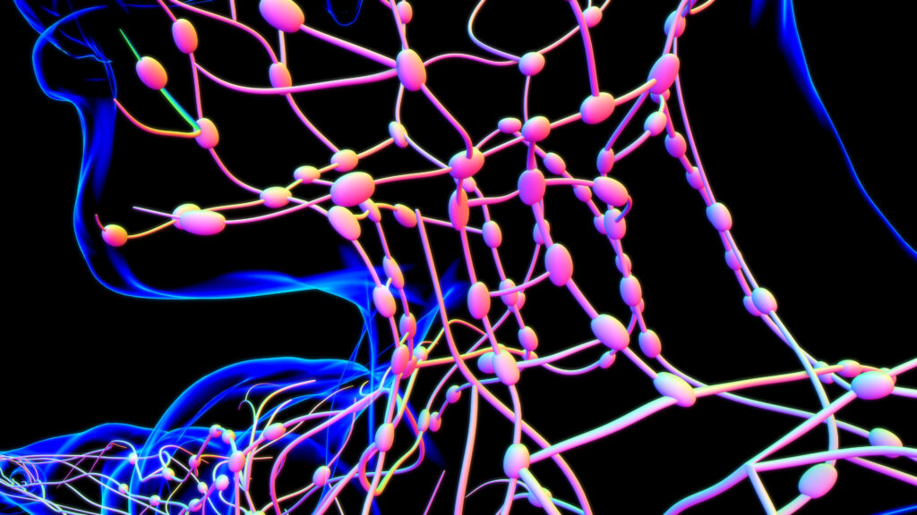
It is generally accepted that prevention is better than cure and the opportunity now exists with modern medical imaging to diagnose disease at an unprecedented early stage. Over the last 10 years there have been enormous improvements in scanning technology with the latest scanners able to visualise the body in incredible detail. However, contrary to what many believe or even claim, it is essential to understand that no single scanner can visualise all parts of the body equally effectively. Different scanners have individual strengths and an experienced clinician knows to choose a specific scanner to diagnose a specific disease.
At Echelon Health we fully recognise this and utilise a variety of the world’s most advanced scanners to provide an overall assessment of those diseases which can lead to early death.
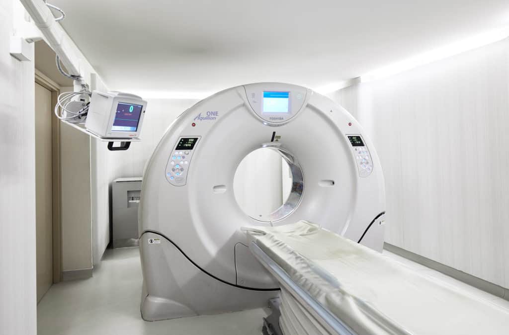
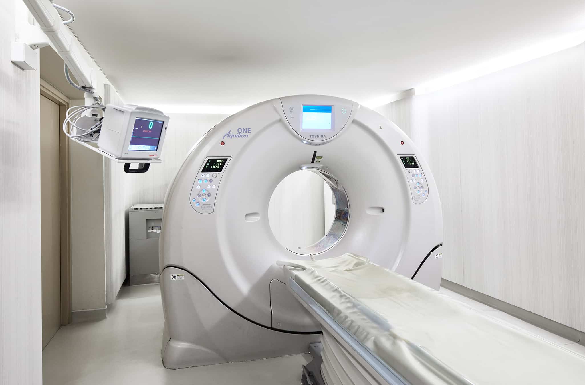
CT scanners
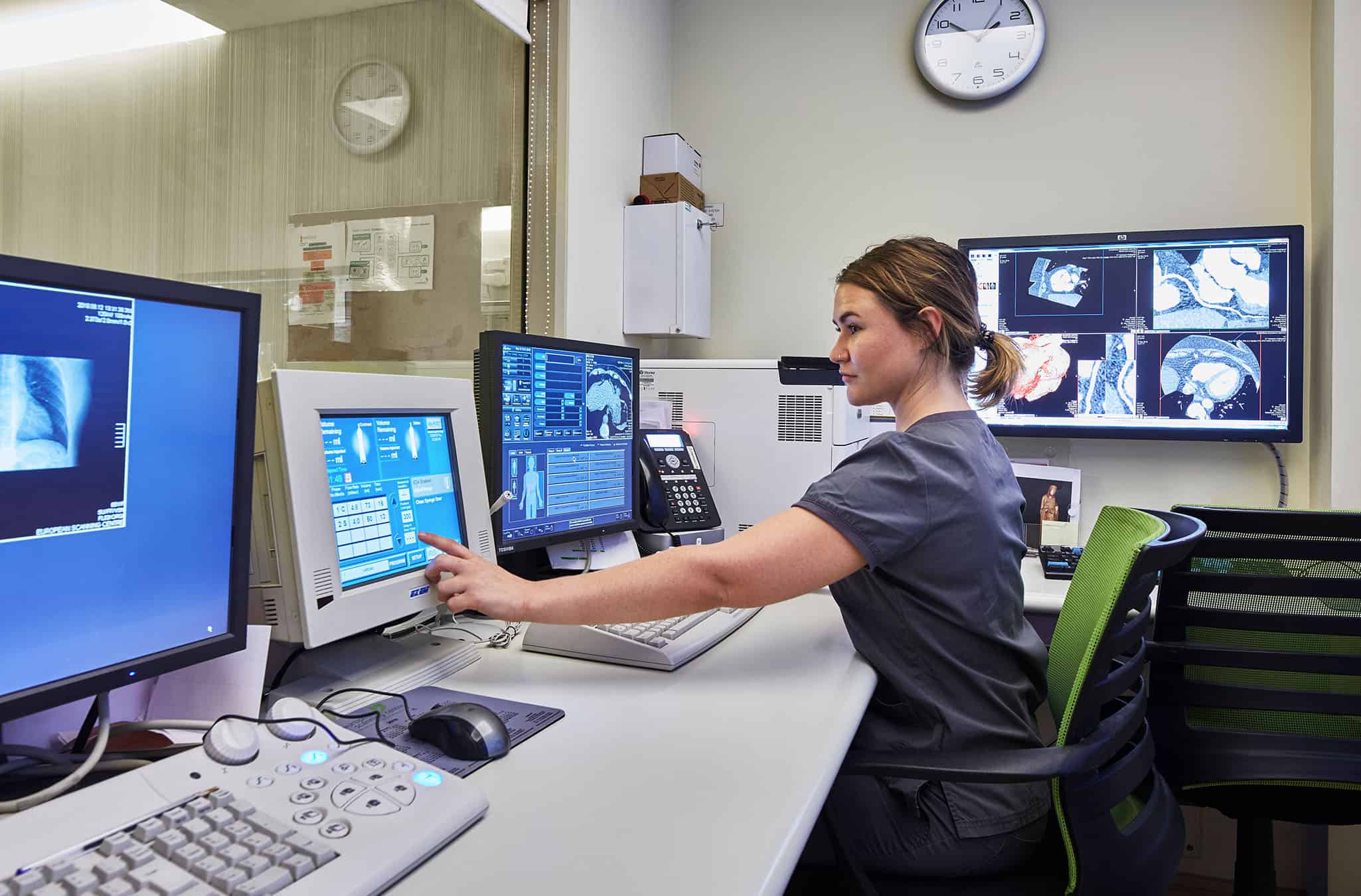
CT scanners are in essence a rotating x-ray tube which revolves around the body at high speed with the resulting x-rays being detected by a series of detectors on the opposite side of the body. These are then converted by powerful computers into high resolution images. Each row of detectors can analyse images from a 0.5mm ‘slice’ of the body and these slices are then combined together to produce an overall image. The original CT scanners used just 1-2 rows of detectors and it took several hours to image the body and 48 hours to analyse the data and produce a picture! The resulting images were understandably very indistinct and blurred. Until recently, the NHS used 16 slice scanners which were a major improvement but were still slow and produced grainy images with significant radiation exposure to patients. In contrast, the Aquilion ONE CT scanner which Echelon Health uses has an incredible 320 detector row which is then mirrored to allow 640 ‘slices’ to be imaged. This allows the largest field of view of any scanner worldwide and in addition to enhancing its accuracy, means the images come at a very low radiation dose.
CT scanners are the only scanner which can adequately image the heart arteries. MRI scanners are unable to visualise the heart arteries in sufficient detail. Echelon Health’s Aquilion ONE CT scanner has a spatial resolution of just 0.3mm and therefore can see inside the arteries in extraordinary detail to determine the earliest signs of any ‘furring up’ (atherosclerosis) which, if left untreated, can lead to a heart attack. CT scanning is also the only means of visualising the so called ‘soft’ plaque which is the most vulnerable type of furring up and that responsible for the majority of causes of sudden death in middle aged men.
CT imaging is also the only means to visualise the lungs in sufficient detail to diagnose early abnormalities. It allows radiologists to detect cancers as small as a couple of millimetres which dramatically increases the chance of subsequent cure. Waiting for any abnormality to become visible on a chest x-ray reduces 5 year survival rates to less than 5%.
Finally, CT is the only means of performing virtual colonoscopy and screening for colorectal cancer – one of the most preventable cancers as almost all of them develop from polyps over many years and which are visible on the colonoscopy.
MRI scanning
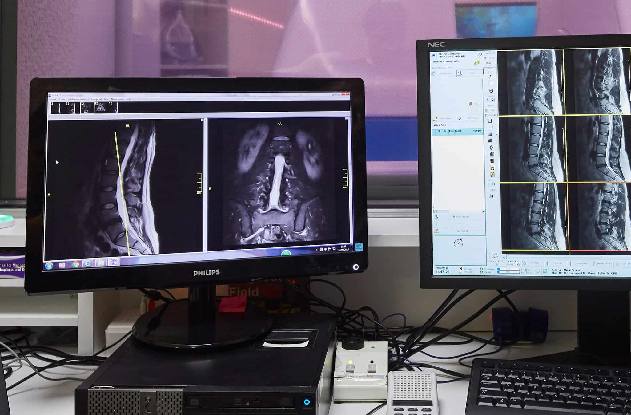
An MRI (magnetic resonance imaging) scanner is a large tube that contains powerful magnets to generate magnetic fields (>30,000 x Earth’s magnetic field) and radio waves to produce images of the inside of the body.
Most of the human body is made up of water molecules, which consist of hydrogen and oxygen atoms. At the centre of each hydrogen atom is an even smaller particle called a proton. Protons are like tiny magnets and are very sensitive to magnetic fields.
When you lie under the powerful scanner magnets, the protons in your body line up in the same direction, in the same way that a magnet can pull the needle of a compass. In the MRI scanner, short bursts of radio waves are then sent to certain areas of the body, knocking the protons out of alignment. When the radio waves are turned off, the protons realign. This sends out radio signals, which are picked up by receivers. These signals provide information about the exact location of the protons in the body. They also help to distinguish between the various types of tissue in the body, because the protons in different types of tissue realign at different speeds and produce distinct signals. In the same way that millions of pixels on a computer screen can create complex pictures, the signals from the millions of protons in the body are combined to create a detailed image of the inside of the body.
MRI scanners are the optimal means of imaging the:
- brain and spinal cord
- bones and joints
- prostate gland
- (occasionally breast)
They cannot adequately image the coronary arteries, lungs or colon.
The strength of the magnets (measured in Tesla) within the MRI scanner largely determines the quality and detail of the scan images. Most MRI scanners, and certainly those within the NHS, generally operate at a strength of 1.5 Tesla which provides a good image quality. However, optimum images are obtained from using a 3 Tesla MRI (twice the normal strength) which provides a greater signal-to-noise ratio. This is especially so for prostate gland imaging and therefore, so called multi-parametric 3T MRI scanning is the new ‘gold standard’ for detecting prostate cancer – the most common cancer in men.
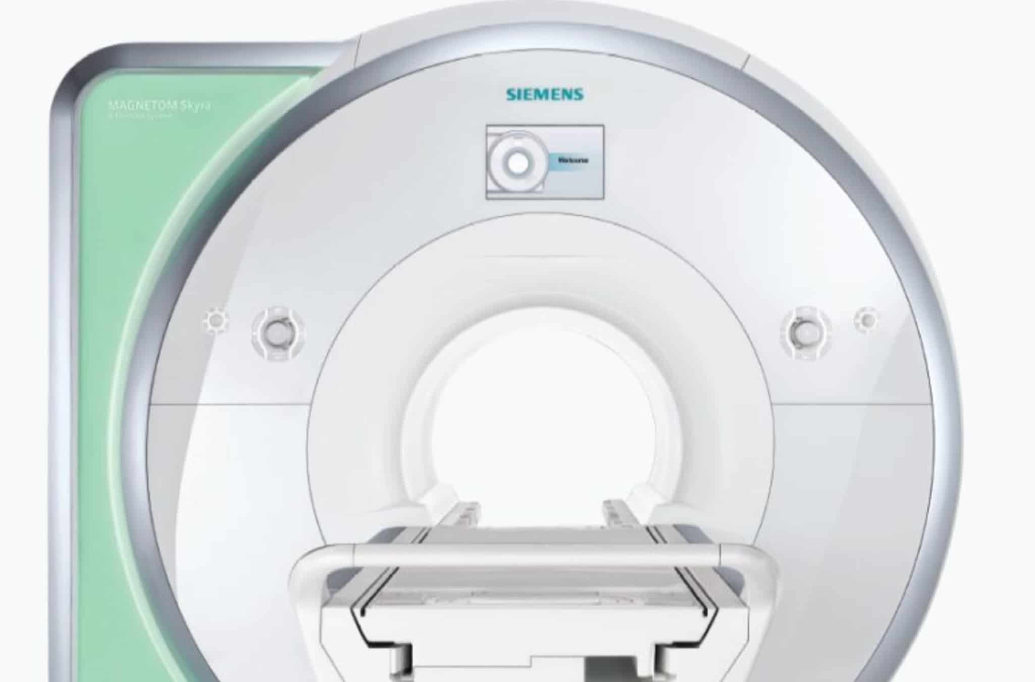
Due to their increased expense and the complexities of operating them, there are currently just a handful of 3T MRI scanners in central London, including one within ESC’s (Echelon Health’s preferred imaging partner) other centre located just a 4 minute walk from our main Harley Street location. We know Echelon Health clients are very happy that we have made the decision to use this specific industry leading scanner rather than using the most convenient and the very short trip for a scanner of this quality and rarity, which delivers significantly better image quality. It is the opinion of our Medical Director that is definitely worth the slight inconvenience to our clients of having to change locations during their assessment.
Ultrasound
An ultrasound scanner utilises high-frequency sound waves to bounce off different parts of the body to create echoes that are detected by the probe and turned into a moving image. This image is displayed on a monitor while the scan is carried out. Different probes are used to image different parts of the body.
Ultrasound scans are optimal for imaging of the thyroid gland, testes and ovaries, and also sometimes of blood vessels in the neck. It can also be used to clarify abnormalities of the liver seen on MRI or CT scan.
While an ultrasound scan is generally quick and painless, the quality of the images depends on both the quality of the technology and the operator. At Echelon Health we use the Canon Aplio ultrasound scanner which is one of the most advanced and complex scanners available with a comprehensive suite of specialist probes to provide optimum imaging of the different body organs. Importantly our scans (with the exception of pelvic transvaginal scans) are performed by senior and very experienced consultant radiologists rather than technicians which increases the accuracy of the reports.
EOS scanner
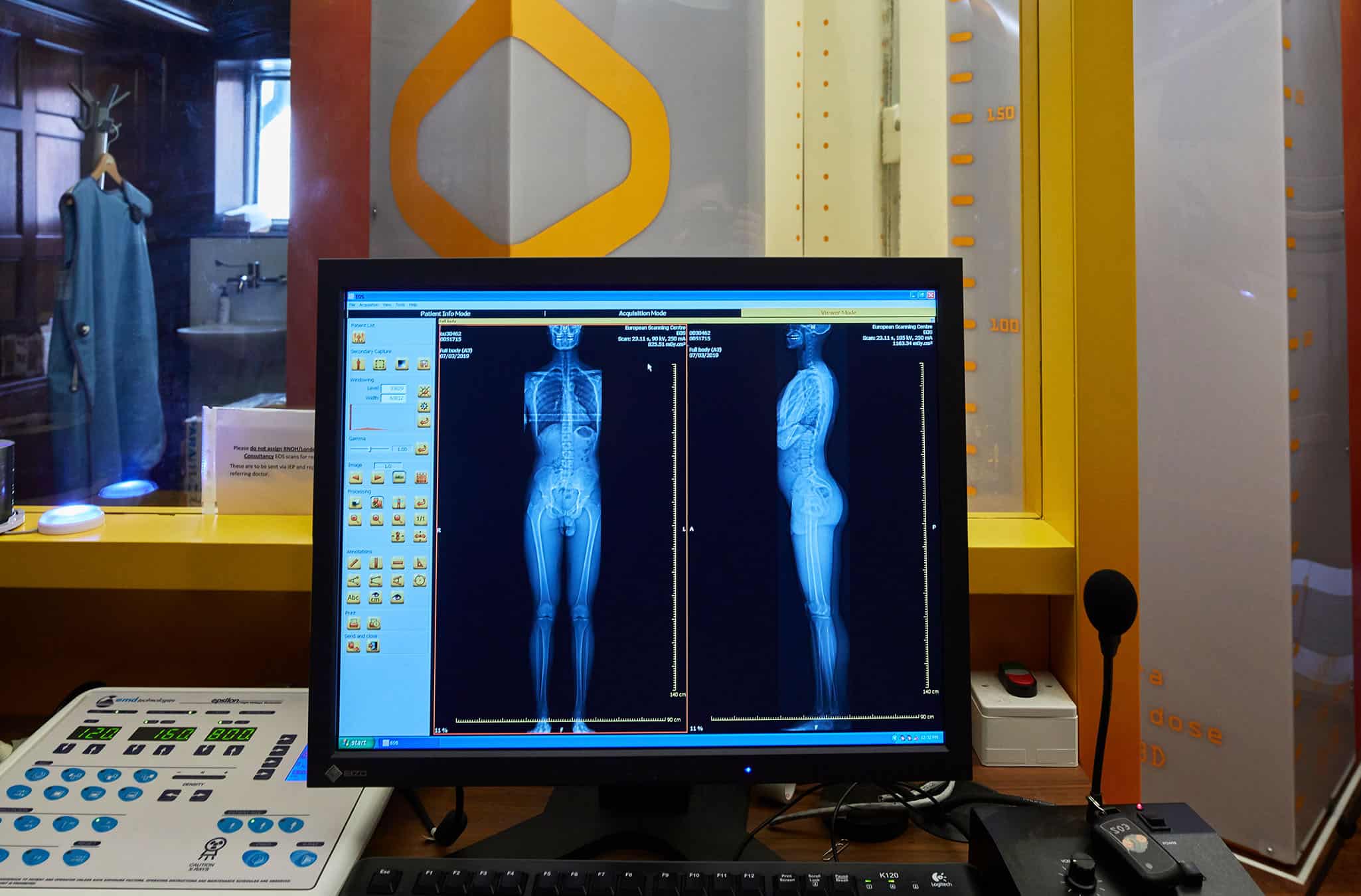
The EOS scanner is a unique upright ultra-low dose CT scanner which utilises Nobel Prize winning detector technology – derived from the CERN project – to enable imaging of the entire skeleton. EOS also allows for 3-D reconstruction of individual bone position, rotation and orientation. This comprehensive view of spine and joint alignment allows for a more accurate assessment of deformities.
Perhaps most importantly is that the scanning occurs in a standing position. All other imaging of the skeleton (with the exception of the ESC upright MRI scanner) requires scanning to be performed in a lying position and therefore does not provide physiological information of what is happening to the skeleton in a real-life weight bearing position. Almost all of us suffer with low back pain at some stage in our lives, which is extremely disabling and can lead to chronic pain and disability. Therefore, having such an accurate assessment of postural and skeletal alignment as well as measurement of leg length provides unrivalled knowledge of predisposing factors and thereby allows for effective preventive strategies to be implemented.
Due to their expense, there are currently just 3 EOS scanners in the whole of the UK, with Echelon Health’s partner ESC having one in our Harley Street centre.
Mammograms
A mammogram is an x-ray image of the breasts used to screen for breast cancer. Mammograms play a key role in early breast cancer detection and help decrease breast cancer deaths.
During a mammogram, the breasts are compressed between two firm surfaces to spread out the breast tissue. Then an x-ray captures black-and-white images of the breasts. Traditionally, these images have been stored on film but recent advances in technology enables them to be stored as digital images. This has been shown to offer several benefits, especially in younger women and those with denser breasts.
- More analysis. Because digital mammograms are stored electronically, they can be analysed by computers as well as by radiologists.
- Easier second opinions. They can easily be sent electronically for analysis. All our mammogram images are reported by 2 separate consultant radiologists.
- Better quality images. The images can be manipulated for better clarity and visibility which is not possible with film mammograms, and this has been shown to significantly increase sensitivity and detection rate of cancers.
- Significantly reduced average radiation dosage. While digital mammograms often take more views of each breast than the film kind they use about 25% less radiation as smaller areas of the breast are imaged in each view.
There are just a handful of centres offering digital screening mammography in London and so we at Echelon Health have selected the one which we regard as offering the best quality breast imaging service with the resulting scans being reported separately by two internationally renowned specialist radiologists. Fortunately, this centre is also located just a few doors away from our Harley Street clinic.
Request a call back